 Students Login
 Teachers Login
The thoracic cavity contains the lungs and heart and is enclosed by the ribs: from the front – the ribs connect to the sternum; at the back – the ribs connect to the spine; upper boundary – clavicle; lower boundary – the diaphragm.

Lesson no. 20
Chest Injuries
The thoracic cavity contains the lungs and heart and is enclosed by the ribs: from the front – the ribs connect to the sternum; at the back – the ribs connect to the spine; upper boundary – clavicle; lower boundary – the diaphragm.
Caused by injury to the thoracic cage.
Apart from severe pain that will affect breathing (the injured person will breathe superficially in response to pain), a rib fracture can be extremely dangerous as it can puncture the lung and cause significant bleeding as a result of injury to arteries and veins located above the rib.

Flail chest is an injury that results from two rib fractures (at least) and in two or more adjacent ribs.
The fragments form a section that is not connected to the rest of the rib cage, which interferes with proper expansion and contraction of the lung. On average, during the first two hours, the muscles around the segment will produce a contraction for the thorax to expand normally during the respiratory process. After two hours the muscles will tire and the segment will float and cause paradoxical breathing. Paradoxical breathing still visually appears to be a proper ascending and descending thorax, except for the floating section that appears to ascend as the thorax descends, and descends as the thorax ascends because it is not connected to the rest of the rib cage.
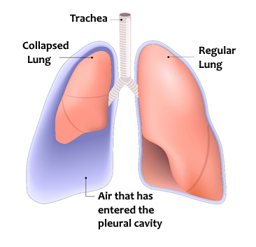
Pneumothorax occurs when air from the lungs or outer space enters the space between the pleural membranes, breaking the pressure between them and detaching the lungs from the inner part of the ribs.
In the inhalation process, the thoracic cage pulls the outer pleural membrane, and it pulls the inner membrane by vacuum pressure (which in turn pulls and expands the lungs). As air enters between the pleural membranes, the vacuum pressure will be eliminated, and the lung will not expand properly.
If the air comes from outside the lung as a result of a penetrating injury, the injury is called an open pneumothorax.
If air comes out of the lung as a result of an inner perforation, the injury is called a closed pneumothorax.
In addition, there is a condition called “Spontaneous Pneumothorax” that occurs most often in tall, lean men who smoke, with a family history, where a small puncture is spontaneously created, and air flows from it into the pleural space.
If it is an open pneumothorax, the signs are:
Treatment that can be given at our level:
In the hospital, the injured person will be treated by having a drain inserted into the pleural space which will drain the air from it.
Like with pneumothorax, and from the new name, we can understand that a hemothorax is formed as blood accumulates between the pleura membranes.
Beyond the severity of the injury to the respiratory tract, as explained with pneumothorax, another danger with hemothorax is massive blood loss, as each side of the thorax can contain about one-third of an adult’s blood volume.
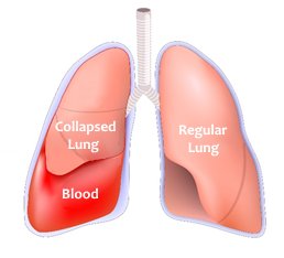
The signs of a blood chest are similar to those of pneumothorax plus signs of shock due to the extensive bleeding:
The treatment provided is the same as the treatment of pneumothorax (although a medic and above can also give an infusion). Hospital treatment is also the same – the insertion of a chest drain to drain the blood from the pleural space.
Tension pneumothorax is created when air enters the pleura, but as a result of the creation of a natural one-way valve, the air is only able to enter the pleura space and is unable to exit (unlike a normal pneumothorax).
The injury means that with each inhalation, more air will enter the inter-pleural space, but upon exhalation, it will not exit, as opposed to a simple pneumothorax. Since the thorax is limited space, the pressure that will accumulate in the inter-pleural space will begin to press against organs near the area – the lung, blood vessels, and the heart.
The pressure pressed against the lung will prevent it from expanding and lead to a condition called lung collapse – atelectasis.
The pressure exerted on the vena cavae (central veins that drain unoxidized blood from the body back into the heart) will decrease the venous return to the heart and thus the heart won’t fill, up to a point where the heart is no longer able to supply blood to the body during systole, a condition called cardiogenic shock.
Tension pneumothorax is a life-threatening condition that requires immediate treatment.
The signs are the same as pneumothorax but more intense:
At this point it is important to say that the injuries can be combined (both air and blood) – namely, hemopneumothorax and tension hemopneumothorax.
