 Students Login
 Teachers Login
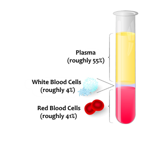
The cardiac system is divided into three parts: pump (heart), pipes (blood vessels) and fluid (blood).

Lesson no. 6
The Cardiovascular System
The cardiac system is divided into three parts: pump (heart), pipes (blood vessels) and fluid (blood).
These consist of three types of blood vessels:
These are usually blood vessels that contain oxygen-saturated blood (except for pulmonary arteries which pump carbon dioxide-saturated blood). It is the most massive blood vessel of them all because it is required to carry the greatest pressure.
It consists of three layers:
Important Arteries:
Coronary arteries: the arteries that nourish the heart muscle itself.
The arteries come from the very beginning of the aorta and engulf the entire heart.
It is important to remember that, unlike the entire circulatory (cardiovascular) system, the heart is nourished by the coronary arteries during diastole as opposed to during systole!
These are usually blood vessels that contain carbon dioxide-saturated blood (except the pulmonary veins that carry oxygen-saturated blood).
Blood Pressure – the pressure exerted by circulating blood against the walls of the blood vessels.
Blood pressure is affected by many factors such as the amount of blood, the heart’s force of contraction, the number of beats per minute, the diameter of blood vessels (which can vary), etc. Blood pressure is divided into systolic pressure (when the rooms of the heart contract) and diastolic pressure (when the rooms of the heart relax).
In a normal adult, systolic blood pressure ranges from 90-140mg and normal diastolic blood pressure ranges from 60-90mg. The number is presented as follows: 120/70.
Proper blood pressure in children and infants (systolic) is calculated as 80 + (age of child multiplied by 2).
Pulse Pressure is defined as the difference between systolic and diastolic blood pressure, where the normal range ranges from 30-60mg.
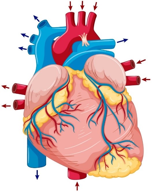
The heart is a muscular organ used to pump blood. Throughout our lives, the heart never stops beating, not even for a moment. This makes the heart muscle very unique. It is roughly the size of a person’s fist, and is located in the center of the thorax, behind the sternum, with a slight tilt to the left.
We’ll look at the heart like a box divided into four chambers:
Divided horizontally – a left side and a right side, with the left side rich in oxygen and the right-side oxygen-depleted.
Divided vertically – atrium and ventricles. At the top of the box are two atria, and at the bottom of the box are two ventricles.
The heart’s muscle (myocardium) is responsible for circulating blood throughout the body and uses a mechanism that utilizes contraction and relaxation.
When the heart contracts, blood flows from the heart to the blood vessels, while during relaxation the heart fills with blood. Between the atrium and ventricles and between the ventricles and large arteries (the aorta and pulmonary artery) are valves that allow blood to flow in only one direction, which keeps the blood volume in the chambers that later circulate to the blood vessels.
The heart consists of three layers:
Around it is the pericardium (or: the heart’s sac) and between them is the pericardial fluid that acts as a lubricant.
First, the atria contract with the ventricles soon following. When the atria are contracted, the ventricles are relaxed and the valves between the atria and ventricles are open and allow blood flow from the atria to the ventricles (diastole). After the blood passes from the atria to the ventricles, the rooms will contract to pump the blood into the large blood vessels, which will circulate blood into the body and lungs. When the ventricles are contracted, the valves between the atria and ventricles are closed, preventing blood flow back to the atrium (systole), as the atria are filled with blood.
The normal adult heart rate is 60-100 beats per minute.
The normal heart rate for a child is 80-120 beats per minute.
The normal heart rate for babies is 100-160 beats per minute.
A heart rate above 100 beats per minute (in an adult) is called tachycardia, while a heart rate below 60 beats per minute (in an adult) is called bradycardia.
We will learn about the cardiovascular structure by following the journey of blood circulation.
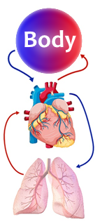
Systemic Circulation: Oxygen-saturated blood found in the left atrium passes through the mitral valve to the left ventricle. From the left ventricle, the blood exits through the aortic valve to the aorta. From the aorta, blood flows to the entire body. Blood flowing through the artery is saturated with oxygen and supplies oxygen to the body’s organs through capillaries. In the capillaries, gases are exchanged with the cells. They pass along oxygen to the cells and receive carbon dioxide back. Carbon dioxide flows from the capillary system and continues to the veins until it reaches the two venae cavae: the superior vena cava which returns blood from the upper part of the body, and the inferior vena cava which flows blood from the lower part of the body back to the heart. The venae cavae enter the right atrium. This blood is oxygen-depleted.
Pulmonary Circulation: Blood saturated in carbon dioxide (CO2) and oxygen-depleted, which is found in the right atrium, flows to the right ventricle through the tricuspid valve. From the right ventricle, the blood flows through the pulmonary valve to the pulmonary artery. The pulmonary artery splits into two and enters the lungs. The lungs then carry out an exchange of gases, where carbon dioxide is exchanged for oxygen. From the lungs, the blood drains into the four pulmonary veins and from there to the left atrium. This blood is oxygen-rich.
Electrical Conduction System: The heart is unable to perform mechanical contraction without electrical stimulation and because of its importance, the heart has its own conduction system.
The electrical current is created in the heart’s main pacemaker (sino-atrial node) which is located in the right atrium. The electric current allows for atrial contraction. After pausing for about a sixth of a second, the current continues to the secondary pacemaker (atrioventricular node), which is positioned at the junction between the atria and the ventricles. The secondary pacemaker transfers the current to the ventricles and causes them to contract. When the heart’s system is functioning properly, the atria will contract before the ventricles, so that blood can fill the ventricles and be ejected into the rest of the circulatory system.
 did you know?
The Cardiovascular System
Heart Disease
Circulatory System
